Chest Muscles Anatomy / 791 Chest Muscles Vectors Royalty Free Vector Chest Muscles Images Depositphotos. Pectoralis major and pectoralis minor. Chest muscle anatomy the pectoralis major muscles also known as the pecs are located on the front of the rib cage and form the major muscles of the chest. The respiratory organs consist of the upper respiratory tract: This mri chest (thorax) axial cross sectional anatomy tool is absolutely free to use. Find the normal cholesterol levels.ways to lower ldl cholesterol.
This is a table of skeletal muscles of the human anatomy. The muscular system is an organ system consisting of skeletal, smooth and cardiac muscles. The pectoralis major and the pectoralis minor, known collectively as your pecs. Four main muscles in the pectoral region exert a force on the upper limb. It's considered to be one of the most effective and reliable methods of measuring muscle activity.
Muscle Masterclass Chest Sebastian Fitness Solutions from www.sebastianfitnesssolutions.com The muscles of the chest and upper back occupy the thoracic region of the body inferior to the neck and superior to the abdominal region and include the muscles of the shoulders. Learn about chest muscle anatomy with free interactive flashcards. These important muscles control many motions that involve moving the arms and head — such as throwing a ball, looking up at the sky, and raising your hand. The muscular system is an organ system consisting of skeletal, smooth and cardiac muscles. (1) the pectoralis major, and (2) the pectoralis minor. Use the mouse scroll wheel to move the images up and down alternatively use the tiny arrows (>>) on both side of the image to move the images.>>) on both side of the image to move the images. The pectoralis major, pectoralis minor, serratus anterior and subclavius. The chest anatomy includes the pectoralis major.
These important muscles control many motions that involve moving the arms and head — such as throwing a ball, looking up at the sky, and raising your hand.
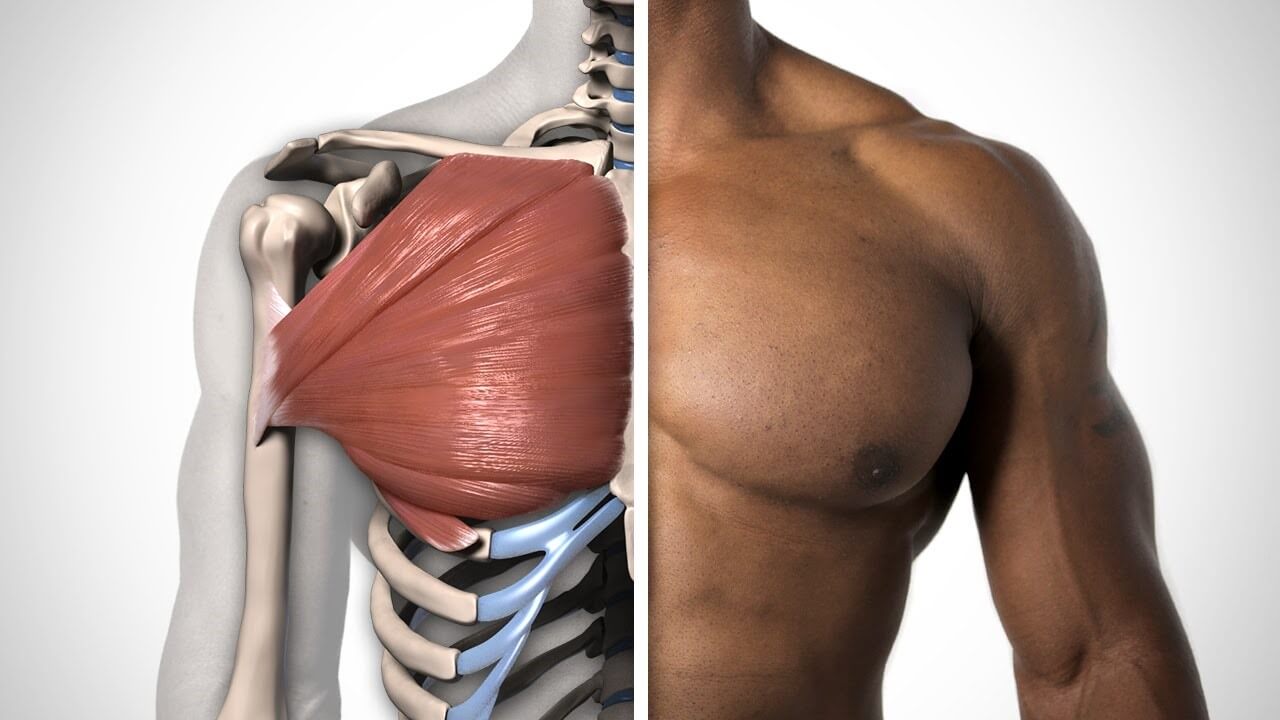
These include pectoralis major, pectoralis minor, serratus anterior, and subclavius. Let's have a detailed look at each of their types and functions. The clavicular head and the sternocostal head. The pectoralis major, pectoralis minor, serratus anterior and subclavius. The clavicular head originates from the front of your collar bone (medial clavicle), then continues down your upper arm bone ( humerus ) where it attaches at the intertubercular sulcus. This mri chest (thorax) axial cross sectional anatomy tool is absolutely free to use. Chest muscles anatomy the chest is made up primarily of two muscles: (1) the pectoralis major, and (2) the pectoralis minor. Muscle anatomy neck 12 photos of the muscle anatomy neck dog neck muscle anatomy, front neck muscle anatomy, muscle anatomy neck, muscle anatomy of neck and shoulder, neck muscle anatomy chart, human muscles, dog neck muscle anatomy, front neck muscle anatomy, muscle anatomy neck, muscle. Educational video describing the muscle anatomy and function of the pectoralis muscles.the human chest consists of two pectoral muscles, the pectoralis major. Pectoralis major and pectoralis minor. Computed tomography (ct) of the chest can detect pathology that may not show up on a conventional chest radiograph (1). To know whether or not an exercise targets the right muscles or not, scientists use a type of test called electromyography (emg).
It contains four muscles that exert a force on the upper limb: Browse 2,552 female chest anatomy stock photos and images available, or start a new search to explore more stock photos and images. The beginner as well as advanced players meet with the problem when building a powerful chest. The pectoralis major, the larger and more superficial, originates at the clavicle (collarbone), the sternum, the ribs, and a tendinous extension of the external oblique abdominal muscle. Let's have a detailed look at each of their types and functions.
Thoracic And Abdominal Muscles Lecturio Online Medical Library from d3uigcfkiiww0g.cloudfront.net Learn about chest muscle anatomy with free interactive flashcards. The clavicular head and the sternocostal head. Chest muscle anatomy the pectoralis major muscles (also known as the pecs) are located on the front of the rib cage, and form the major muscles of the chest. These important muscles control many motions that involve moving the arms and head — such as throwing a ball, looking up at the sky, and raising your hand. Find the normal cholesterol levels.ways to lower ldl cholesterol. Educational video describing the muscle anatomy and function of the pectoralis muscles.the human chest consists of two pectoral muscles, the pectoralis major. The muscle has two heads: Muscle anatomy neck 12 photos of the muscle anatomy neck dog neck muscle anatomy, front neck muscle anatomy, muscle anatomy neck, muscle anatomy of neck and shoulder, neck muscle anatomy chart, human muscles, dog neck muscle anatomy, front neck muscle anatomy, muscle anatomy neck, muscle.
Learn about each of these muscles, their locations, functional anatomy and exercises for them.
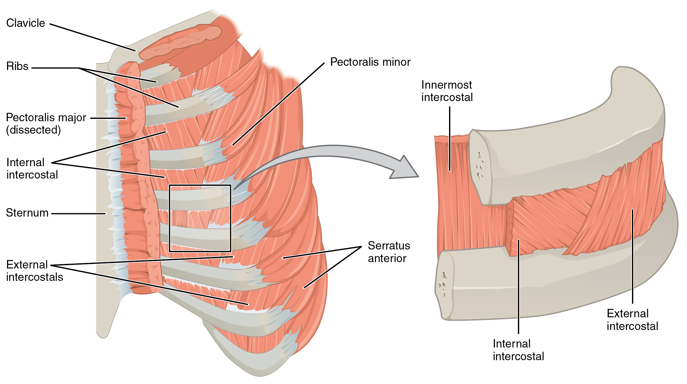
The respiratory organs consist of the upper respiratory tract: Neuromuscular therapy muscle diagram latissimus dorsi male chest trigger point therapy chest muscles muscle anatomy beauty first anatomy arm diagram human lasalle muscles sakart. Not everyone, however, has the chance to achieve stunning results when working on chest muscles. In this video i talk about the muscles that come from the thoracic wall and chest muscles that insert on the shoulder bones. There are two such muscles on each side of the sternum (breastbone) in the human body: The pectoralis major, pectoralis minor, serratus anterior and subclavius. Beneath the pectoralis major is the pectoralis minor, a thin, triangular muscle. Browse 2,552 female chest anatomy stock photos and images available, or start a new search to explore more stock photos and images. Muscles of the chest and their functions you have two mighty muscles on both sides of your chest: It's considered to be one of the most effective and reliable methods of measuring muscle activity. Muscles the dominant muscle in the upper chest is the pectoralis major. Educational video describing the muscle anatomy and function of the pectoralis muscles.the human chest consists of two pectoral muscles, the pectoralis major. These include pectoralis major, pectoralis minor, serratus anterior, and subclavius.
The chest anatomy includes the pectoralis major. Chest muscle anatomy the pectoralis major muscles also known as the pecs are located on the front of the rib cage and form the major muscles of the chest. The chest or thorax is the region between the neck and diaphragm that encloses organs, such as the heart, lungs, esophagus, trachea, and thoracic diaphragm. The muscular system is an organ system consisting of skeletal, smooth and cardiac muscles. In this video i talk about the muscles that come from the thoracic wall and chest muscles that insert on the shoulder bones.
Amazon Com Anatomy Torso Chest Muscle Print Sra3 12x18 Conqueror Laid Paper Handmade from images-na.ssl-images-amazon.com In this video i talk about the muscles that come from the thoracic wall and chest muscles that insert on the shoulder bones. (1) the pectoralis major, and (2) the pectoralis minor. There are two such muscles on each side of the sternum (breastbone) in the human body: These important muscles control many motions that involve moving the arms and head — such as throwing a ball, looking up at the sky, and raising your hand. The pectoral region is located on the anterior chest wall. Neuromuscular therapy muscle diagram latissimus dorsi male chest trigger point therapy chest muscles muscle anatomy beauty first anatomy arm diagram human lasalle muscles sakart. Chest muscle anatomy the pectoralis major muscles also known as the pecs are located on the front of the rib cage and form the major muscles of the chest. Anatomy chart courtesy of fcit the pecs attach to the humerus near the shoulder joint and originate on the breastbone in the center of the chest.
External pectoral muscles the external chest muscles include:
Not everyone, however, has the chance to achieve stunning results when working on chest muscles. Start with a pair of dumbbells extended above your chest. Let's have a detailed look at each of their types and functions. All about the chest muscles the chest anatomy includes the pectoralis major, pectoralis minor and the serratus anterior. Learn about each of these muscles, their locations, functional anatomy, and exercises for them. O muscles—sternocleidomastoid, anterior and middle scalene, infrahyoid, pectoralis major and o diaphragm. Beneath the pectoralis major is the pectoralis minor, a thin, triangular muscle. Neuromuscular therapy muscle diagram latissimus dorsi male chest trigger point therapy chest muscles muscle anatomy beauty first anatomy arm diagram human lasalle muscles sakart. (1) the pectoralis major, and (2) the pectoralis minor. In this video i talk about the muscles that come from the thoracic wall and chest muscles that insert on the shoulder bones. The muscle has two heads: Pectoralis major and pectoralis minor. It contains four muscles that exert a force on the upper limb:
Share :
Post a Comment
for "Chest Muscles Anatomy / 791 Chest Muscles Vectors Royalty Free Vector Chest Muscles Images Depositphotos"


Post a Comment for "Chest Muscles Anatomy / 791 Chest Muscles Vectors Royalty Free Vector Chest Muscles Images Depositphotos"